Friday Factoid: The Tryptophan In Your Turkey: What You Didn’t Know

Tryptophan found in turkey is believed to be the legendary reason why people always doze off for little naps on Thanksgiving Day.  In fact, “Tryptophan is an amino acid that can be found in several foods, which include dairy products, soy products, seafood, poultry and beans” (BeneFit from: Tryptophan, 2008) and there is even more tryptophan in cheese and chicken breast than there is in turkey, according to Elder (2009). To debunk the myth, Elder (2009) says, there is not enough tryptophan in your Thanksgiving turkey to tire you out. However, the tryptophan in your turkey is a precursor to calming, feel-good serotonin.


It seems tryptophan in our food is linked to serotonin, and melatonin. Thornton and Whitley (2012) confirmed the synthesis of serotonin and melatonin can be controlled by tryptophan ingestion (p. 40). Interestingly, Esteban, Nicolaus, Garmundi, Rial, Rodríguez, Ortega, EIbars, (2004) found differences in tryptophan  ingestion at  the beginning of light or dark phases in rats (p. 41). “The administration of  L-tryptophan during the light time  increased the brain synthesis and metabolism of serotonin. However, at night, tryptophan’s administration led to a smaller increase in the synthesis of serotonin than by day, although the turnover remained  unchanged,  suggesting that,  in the dark phase, serotonin is used as a substrate for melatonin synthesis” (Thornton & Whitley, 2012, p. 40).  Esteban et  al., (2004) results imply, “The  difference  between  the effects of increased tryptophan intake during light and dark phases suggests that  tryptophan  hydroxylase  activity  presents  circadian  fluctuations  which seem to be clock controlled” (p. 41).  So, it seems that the legend behind the Thanksgiving naps can in some ways be linked to tryptophan, and tied to our circadian rhythms.


Tryptophan due to its connection to serotonin has been somewhat studied with its role in depression. Parker and Brotchie, (2011) revealed, “There is limited evidence suggesting that depressed individuals, especially those with a melancholic depression, have decreased tryptophan levels. However, results showing a causal contribution or are a consequence of a depressed state remains an open question. Neither the less, the researchers support there is a small database claiming tryptophan preparations benefit people with depressed mood states.”


In conclusion, turkey has tryptophan but other food such as cheese and chicken breast have higher quantities of this amino acid. The amount of tryptophan you eat on Thanksgiving from turkey is not necessarily enough to make you tired, but it could have an impact on your circadian rhythm. The tryptophan you consume impacts your serotonin, and melatonin, which is likely to impact your mood. So therefore, Have A Great Increase of Serotonin on Your Thanksgiving!


P.S. According to The 10 Foods For A Good Night’s Sleep, “Tryptophan works when your stomach is basically empty, not overstuffed, full of protein and not carbohydrates.”


HAPPY THANKSGIVING!!!


References

BeneFit from: Tryptophan. (2008). Cycling Weekly, 32.


Elder, N. (2009). The Question: Does Turkey Make You Sleepy?. Bon Appetit, 54(11), 47.


Esteban, S., Nicolaus, C., Garmundi, A., Rial, R.V., Rodríguez, A., Ortega, EIbars, C. B. (2004). Effect  of orally administered  L-tryptophan on  serotonin, melatonin and  the  innate  immune  response. Molecular and Cellular Biochemistry, 267, 39-46.


Parker, G., & Brotchie, H. (2011). Mood effects of the amino acids tryptophan and tyrosine. Acta             Psychiatrica Scandinavica, 124(6), 417-426. doi:10.1111/j.1600-0447.2011.01706.x

The 10 Foods for a Good Night’s Sleep. (2007). Office Solutions, 24(2), 9.


Thornton, S. H., & Whitley, B. L. (2012). Tryptophan : Dietary Sources, Functions and Health Benefits. New York: Nova Science Publishers, Inc.


Katy Roth, M.A., CRC

WKPIC Doctoral Intern

Friday Factoids Catch-Up: How Biological Processes Impact Sleep


There are many factors that contribute to sleep deprivation for example bathroom trips, sleep schedules, temperature, noise, and technological devices. However, there are biological processes that impact sleep as well. Specifically, there are three biological processes that are controlled through involvement of the brainstem, and two divisions of the hypothalamus, which are the anterior hypothalamus and the suprachiasmatic nuclei (SCN).


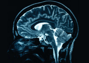
First, the brainstem plays a vital role in REM (rapid eye movement) and NREM (non- rapid eye movement). “The brainstem controls events of REM sleep” Pinel, 2010, pg. 364). REM occurs under the eyelids and was discovered in the 1950’s (Pinel, 2010, pg. 343). Reinoso-Suárez, de Andrés, Rodrigo-Angulo and Garzón (2001) found, “The ventral part of the oral pontine reticular nucleus (vRPO) is a demonstrated site of the brainstem REM sleep-wake cycle, as well as with other brain components responsible for the production of different occurrences related to REM sleep.” Non-REM sleep (NREM) is referred to as slow wave sleep (de Andrés, Garzón, & Reinoso-Suárez, 2011). NREM is vital for standard physical and intellectual functioning and behavior (de Andrés, Garzón, & Reinoso-Suárez, 2011). Further, Villablanca (2004) stated, “Waking can occur independently in both the forebrain and brainstem, but true NREM and REM sleep producing mechanisms exist entirely in the forebrain and brainstem.”


Secondly, the hypothalamus plays a key role in sleep. Specifically, the anterior hypothalamus and adjacent basal forebrain are thought to promote sleep (Pinel, 2010, pg. 355). The anterior hypothalamus is in the basal-forebrain area. “Activation and deactivation of certain cells in the hypothalamus shuts off the arousal system during sleep. Other hypothalamic neurons stabilize the activation and deactivation, however if the switching of cells/neurons is absent this results in inappropriate sleep occurrences, such as disorders like narcolepsy” (Saper, Scammell & Lu, 2005).


From literature the suprachiasmatic nuclei (SCN) in the hypothalamus plays a role in sleep also. “The suprachiasmatic nuclei is situated bilaterally in the hypothalamus, just above the optic chiasm” (Hobson & Pace-Schott, 2002). The SCN is composed of two major subdivisions, the core and the shell. “The core region of the SCN obtains information about the daily light cycle through the retinohypothalamic tract (RHT)” (Takahashi, Hee-Kyung, Ko & McDearmon, 2008). “Neurons in the SCN core correspond with the rhythmic SCN shell. Cells in the rhythmic SCN shell comprise molecular clocks driven by an autoregulatory transcription translation loop” (Antle & Silver, 2005). The SCN controls circadian rhythms (also known as the circadian clock). Interestingly, “Circadian rhythms govern a variety of biological processes in living systems, stretching from bacteria to humans” (Takahashi, Hee-Kyung, Ko & McDearmon, 2008). The suprachiasmatic nucleus of the mammalian hypothalamus contains a circadian clock for timing of diverse neuronal, endocrine, and behavioral rhythms, such as the cycle of sleep and wakefulness (Sakai, 2014). The timing mechanisms of the SCN are dependent on the firing patterns of SCN neurons. During the night SCN neurons tend to be inactive, start to fire at dawn, and fire at a leisurely stable pace all day (Pinel, 2010, pg. 354). Importantly, it seems as though genetics also influences the SCN. Hobson and Pace-Schott (2002) stated, “The molecular circadian clock is genetically controlled and synchronously expressed holistically and individually by 20,000 cells in the mammalian hypothalamus.”


While it is important to be mindful of the many factors in the sleep environment that may impact how much sleep we get, and how rested we feel, there are also biological processes located in our brain, as well as genetics to some degree which impact sleep. It seems our brain has a major role in REM sleep, NREM sleep, and our natural Circadian rhythm as well.


References

Antle, M. C., & Silver, R. (2005). Orchestrating time: arrangements of the brain circadian clock. Trends in neurosciences, 28(3), 145-151.


de Andrés, I., Garzón, M., & Reinoso-Suárez, F. (2011). Functional anatomy of non-REM sleep. Frontiers in Neurology, 2, 70. doi:10.3389/fneur.2011.00070

Hobson, J. A., & Pace-Schott, E. F. (2002). The Neurobiology of Sleep: Genetics, cellular physiology and subcortical networks. Nature Reviews Neuroscience,      3, 591.


Pinel, John  P.J. (2010). Biopsychology, Ninth Edition.  Pearson Education, Inc.


Reinoso-Suárez, F., de Andrés, I., Rodrigo-Angulo, M. L., & Garzón, M. (2001). Brain structures and mechanisms involved in the generation of REM sleep. Sleep medicine reviews, 5, 63-77.


Sakai, K. (2014). Single unit activity of the suprachiasmatic nucleus and surrounding neurons during the wake–sleep cycle in mice. Neuroscience, 260, 249-264.


Saper, C. B., Scammell, T. E., & Lu, J. (2005). Hypothalamic regulation of sleep and circadian rhythms. Nature, 437, 1257-1263.


Takahashi, J. S., Hee-Kyung, H., Ko, C. H., & McDearmon, E. L. (2008). The genetics of mammalian circadian order and disorder: implications for physiology and disease. Nature Reviews Genetics, 9(10), 764-775. doi:10.1038/nrg2430


Villablanca, J. R. (2004). Counterpointing the functional role of the forebrain and of the brainstem in the control of the sleep–waking system. Journal Of Sleep Research, 13, 179-208. doi:10.1111/j.1365-2869.2004.00412.x


Katy Roth, M.A., CRC

WKPIC Doctoral Intern


Friday Factoids: They Creep, They Crawl: Our Fear of Snakes and Spiders


With Halloween right around the corner, it’s the perfect time to talk about the things that go bump in the night, the things that send shivers down our spine, and the things that slither and crawl. Most people have a fear of snakes and spiders (Hoel, Hellmer, Johansson, & Gredebäck, 2017). Just the sight of one of these creepy critters can send people running, but is this fear learned or instinctual?


Many of our fears are learned, however, others are innate (Leahy, 2008). For example, eating bad fish can cause us to become ill, and we may learn to avoid similar fish (Leahy, 2008). This learned aversion is the result of one-trial learning. Yet, we have many fears that did not require trial learning. Most of us are born with a fear of heights, yet we don’t have to fall from a great height to know that heights scare us (Leahy, 2008). Instead, this fear is instinctual and useful in protecting us from potential harm.


In regards to snakes and spiders, prior research had difficulty determining if the fear was learned from parents and others in the environment or an instinctual fear (Max Planck Institute for Human Cognitive and Brain Sciences [MPIHCBS], 2017). Others had determined that most people, even those who lived in cities with no exposure to these creatures, had a deep fear of snakes and spiders, yet past research looked at adults and young children who might have learned this fear from parents or grandparents exposed to these tiny terrors (MPIHCBS, 2017). A new study took a different approach and showed infants pictures of flowers paired with spiders and fish paired with snakes (Hoehl et al., 2017). When examining the snakes and spiders, infants’ pupils demonstrated an increased dilation when compared to their neutral pairings, suggesting a sympathetic response to these frightening stimuli (Hoehl et al., 2017). These findings suggest that our fear of snakes and spiders, much like our fear of heights, is instinctual and meant to help us avoid potential threats (Hoehl et al., 2017).


References:
Hoehl, S., Hellmer, K., Johansson, M., & Gredebäck, G. (2017). Itsy bitsy spider…: Infants react with increased arousal to spiders and snakes. Frontiers in Psychology, 8. doi: 10.3389/fpsyg.2017.01710


Leahy, R.L. (2008). Are we born to be afraid? Psychology Today. Retrieved from https://www.psychologytoday.com/blog/anxiety-files/200805/are-we-born-be-afraid


Max Planck Institute for Human Cognitive and Brain Sciences. (2017). Itsy bitsy spider: Fear of spiders and snakes is deeply embedded in us. ScienceDaily. Retrieved from www.sciencedaily.com/releases/2017/10/171019110953.htm


Michael Daniel, MA
WKPIC Doctoral Intern


Friday Factoids: How Fathers See Their Babies


Emotions are “contagious”, especially for babies (American Psychological Association, 2017; Waters, West, Karnilowicz, & Mendes, 2017).  When mothers hold their babies, the mother’s affect is often “caught” by the baby.  This emotional transmission can be seen when mothers are in a high-anxiety situation, the baby exhibits a sympathetic response; however, in low-stress scenarios, the baby demonstrates a parasympathetic response (Waters et al., 2017).


Past research has shown that mothers and fathers frequently respond differently to boy and girl babies (APA, 2017; Mascaro, Rentscher, Hackett, Mehl, & Rilling, 2017).  Parents often talk more to girls yet restrict their behaviors, while boys are talked to less but allowed to engage in more risk-taking activities (e.g. climbing, “rough housing”).  New research has taken another step and examined the neural functioning of fathers when with their children (Mascaro et al., 2017).  When viewing pictures of their daughters smiling, fathers demonstrate a response in the orbitofrontal cortex, yet fathers experience the same response when viewing their sons exhibiting a neutral response (Mascaro et al., 2017).  These neural responses suggest fathers have differing expectations for their children.  Through the affect contagion scenario, it might be hypothesized that our babies are being primed early in how they express emotions.  Girls are likely learning that an affective response is positive, while boys may be learning that a restricted range of emotions is desirable.


References:

American Psychological Association. (2017). Parent-child interactions.  Particularly Exciting Experiments in Psychology, 98. Retrieved from http://www.apa.org/pubs/highlights/peeps/issue-98.aspx

Mascaro, J. S., Rentscher, K. E., Hackett, P. D., Mehl, M. R., & Rilling, J. K. (2017). Child gender influences paternal behavior, language, and brain function. Behavioral Neuroscience, 131(3), 262–273. http://dx.doi.org/10.1037/bne0000199

Waters, S. F., West, T. V., Karnilowicz, H. R., & Mendes, W. B. (2017). Affect contagion between mothers and infants: Examining valence and touch. Journal of Experimental Psychology: General, 146(7), 1043–1051. http://dx.doi.org/10.1037/xge0000322


Michael Daniel, MA
WKPIC Doctoral Intern


Friday Factoids: Your Brain on Glycine


Until recently, research has suggested early psychosis has its roots in poor glycine production (Elsevier, 2017).  This hypothesis was due, in part, to evidence that psychosis was caused by impaired NMDA receptors, glutamate-gated channels responsible for numerous biological functions as well as leaning, memory, and neuroplasticity (Blake and VanDongen, 2009; Elsevier, 2017).  It was believed that supplying patients with additional glycine would alleviate symptoms of early psychosis; however, drugs targeting NMDA receptors have demonstrated limited success in the past (Blanke and VanDongen, 2009; Elsevier, 2017).


New research from Kim et al. (2017) suggests that first-episode psychosis is not linked to a deficit of glycine but a surplus.  While this finding does not discredit the NMDA receptor hypofunction hypothesis, it does create new questions.  Future research will need to examine if accumulation of glycine is due to reduced glycine use or overproduction of the amino acid (Kim et al., 2017).  Regardless, the new finding helps spread light on why glycine treatment has demonstrated limited effectiveness in the past, and may warrant new avenues of treatment for first-episode psychotic symptoms.


References:
Blanke, M.L. & VanDongen, A.M. (2009). Activation mechanisms of the NMDA receptor. In A.M. VanDongen (Ed.), Biology of the NMDA Receptor. Boca Raton, FL: CRC Press/Taylor & Francis.


Elsevier. (2017). Brain chemical abnormalities in earliest stage of psychosis identified. ScienceDaily. Retrieved October 12, 2017 from www.sciencedaily.com/releases/2017/10/171011120339.htm


Kim, S., Kaufman, M., Cohen, B.M., Jensen, J.E., Coyle, J.T., Du, F., & Öngür, D. (2017). In vivo brain glycine and glutamate concentrations in patients with first-episode psychosis measured by echo-time-averaged proton magnetic resonance spectroscopy at 4T. Biological Psychiatry. doi: http://dx.doi.org/10.1016/j.biopsych.2017.08.022


Michael Daniel, MA
WKPIC Doctoral Intern